When I tell an owner their German Shepherd has hip dysplasia, I watch their face fall. The diagnosis sounds devastating, and the internet has not helped with its apocalyptic descriptions. But hip dysplasia is not a death sentence. It is not even necessarily a sentence to chronic pain. What it is, fundamentally, is a developmental abnormality that we can manage, treat, and in many cases largely overcome.
Understanding what hip dysplasia actually is, rather than what marketing materials and worried forum posts claim it to be, is the first step toward helping your dog. So let me explain this condition the way I explain it to clients in my office, with the anatomical details that matter and without the unnecessary alarm.
The Normal Canine Hip
To understand what goes wrong in dysplasia, you need to understand what a healthy hip looks like. The canine hip is a ball-and-socket joint, one of the most elegant mechanical designs in the mammalian body. The ball is the femoral head, a smooth spherical structure at the top of the thigh bone. The socket is the acetabulum, a cup-shaped depression in the pelvis.
In a normal hip, the femoral head fits snugly into the acetabulum. The coverage is deep and complete. Articular cartilage, a remarkably smooth tissue with a coefficient of friction lower than ice on ice, lines both surfaces. Synovial fluid lubricates the joint. A fibrous capsule and powerful muscles hold everything in place while allowing smooth, pain-free movement through a wide range of motion.
When your dog runs, jumps, or simply walks across the room, forces equivalent to several times their body weight pass through this joint. A healthy hip handles this load for a decade or more without complaint. The design works beautifully when everything develops correctly.
What Goes Wrong in Dysplasia
Hip dysplasia is fundamentally a problem of fit. The ball and socket develop abnormally, resulting in a joint where the components do not match properly. This mismatch can take several forms:
Shallow acetabulum: The most common abnormality is insufficient socket depth. Instead of cupping the femoral head securely, the acetabulum provides only partial coverage. The femoral head sits high and loose, like a tennis ball balanced on a saucer rather than nestled in a cup.
Femoral head abnormalities: The ball itself may develop with an abnormal shape. Instead of a smooth sphere, it becomes flattened or irregular. This further compromises the articulation and accelerates wear.
Joint laxity: Even when the bony structures appear reasonably normal on radiographs, the ligaments and capsule may be too loose, allowing excessive movement. This laxity causes the femoral head to subluxate, partially slipping out of the acetabulum during normal activity.
The critical point is that dysplasia is a developmental condition. Puppies are not born with arthritic hips. They are born with genetic predispositions and structural tendencies that, combined with environmental factors during growth, result in abnormal joint development. By the time skeletal maturity is reached, the damage is done.
The Cascade of Consequences
A dysplastic hip might function reasonably well in a young dog. The cartilage is still intact, the muscles are strong, and the dog compensates without apparent difficulty. But the mechanical mismatch creates problems that compound over time.
Cartilage damage: When the femoral head does not seat properly, forces concentrate on small contact areas instead of distributing across the entire joint surface. This focal overloading damages the articular cartilage, which has almost no capacity for self-repair. Once damaged, cartilage degrades progressively.
Inflammation: Damaged cartilage releases inflammatory mediators into the joint fluid. This chronic low-grade inflammation further damages the remaining cartilage, creating a self-perpetuating cycle of destruction.
Osteoarthritis: As cartilage erodes, the underlying bone responds by forming new bone at the joint margins. These osteophytes are the body's attempt to stabilize an unstable joint, but they also cause mechanical impingement and pain. Eventually, bone grinds on bone where smooth cartilage once provided frictionless motion.
Muscle atrophy: Dogs naturally protect painful joints by using them less. Over time, the muscles supporting the affected hip weaken from disuse. This further destabilizes the joint and shifts more load to other structures, often causing secondary problems in the spine or opposite limb. Low-impact activities like structured swimming therapy can counteract this atrophy without adding stress to the compromised joint.
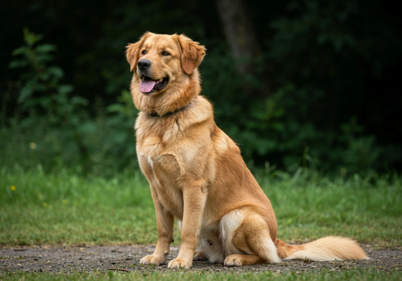
Why Shepherd Breeds Are So Affected
German Shepherds and other herding breeds are disproportionately affected by hip dysplasia. This is not coincidence but the result of breeding history and structural characteristics.
The German Shepherd's distinctive rear angulation, the sloped topline and exaggerated hock angles that define the show ring standard, place unusual biomechanical demands on the hip joint. Working-line dogs with more moderate structure show lower dysplasia rates than heavily angulated show-line dogs, suggesting that conformation itself contributes to the problem.
Additionally, the breed's popularity has led to widespread production by breeders who prioritize appearance or profit over health testing. The genetic factors underlying hip dysplasia have been amplified rather than selected against in many breeding programs.
The Signs You Might Notice
Hip dysplasia can present differently depending on the dog's age, severity of the condition, and individual pain tolerance. Some common signs include:
- Bunny hopping: Using both rear legs together when running rather than alternating normally. This gait pattern reduces stress on individual hip joints.
- Difficulty rising: Stiffness after rest, particularly first thing in the morning or after long naps. The dog may need multiple attempts to stand.
- Reluctance to climb: Avoiding stairs, reluctance to jump into vehicles, or hesitation before getting on furniture.
- Decreased activity: Loss of interest in play, shorter walks, or reduced enthusiasm for previously enjoyed activities.
- Narrow stance: Standing with the rear legs close together to shift weight toward the center of gravity.
- Muscle changes: Loss of muscle mass over the hips and thighs, sometimes with compensatory enlargement of the shoulder muscles.
- Audible clicking: Some dysplastic hips produce sounds during movement as the femoral head subluxates and reduces.
The Importance of Early Detection
I cannot emphasize enough the value of early detection. A puppy identified with lax hips at four months has options that disappear by eighteen months. Surgical interventions like juvenile pubic symphysiodesis or triple pelvic osteotomy can only be performed before skeletal maturity.
Even when early surgical intervention is not pursued, knowing your puppy has hip laxity allows you to modify exercise, optimize nutrition, and maintain ideal body weight during the critical growth period. These environmental modifications can significantly influence how severely the condition manifests.
For adult dogs, early recognition of developing problems allows for conservative management before arthritis becomes severe. Starting appropriate therapy when changes are mild yields much better long-term outcomes than waiting until the dog is obviously crippled.
Diagnosis: More Than Just an X-Ray
Proper diagnosis of hip dysplasia involves clinical examination and imaging. The physical exam includes gait observation, palpation of muscle mass, range of motion testing, and specific manipulative tests like the Ortolani test that assess joint laxity.
Radiographs remain the gold standard for visualization. Standard hip-extended views, as used by the OFA evaluation system, show joint conformation and secondary changes. Distraction radiographs, as used by PennHIP, quantify joint laxity more precisely.
Neither clinical examination nor imaging alone tells the complete story. I have seen dogs with terrible-looking radiographs move comfortably and dogs with mild changes on film show significant pain. The diagnosis integrates all available information with the understanding that our goal is helping the individual dog in front of us, not treating an X-ray.
Looking Forward
A hip dysplasia diagnosis is not the end of the road. Depending on your dog's age, severity of changes, and your circumstances, options range from conservative management to surgical intervention. Many dysplastic dogs live long, active, comfortable lives with appropriate care.
What matters most is accurate understanding, realistic expectations, and committed management. The dogs I see struggle are not necessarily those with the worst hips. They are the ones whose owners were misinformed, delayed seeking help, or expected miracles from unproven treatments. Honest assessment and practical intervention give dysplastic dogs their best chance.
In the following articles on this site, I will walk you through everything from screening and diagnosis to treatment options and long-term management. Hip dysplasia is a challenge, but it is one we can meet together.